Anatomy Of The Upper Chest Area : Zyfbiwlyd7hzym : There are 8 spinal nerves that originate from the cervical spine.. Anatomy of the chest and shoulder, anatomy of the chest organs, anatomy of the chest wall, anatomy of the chest wall and pleura, anatomy of upper chest area, human. Here is a list of possible conditions a doctor might diagnose as causes of upper. What is a pain doctor? Anatomy of the upper chest area / anatomy of the upper chest area / anatomy of the mid to. The upper part of the chest, known as the pectoralis major clavicular head, is one of the.
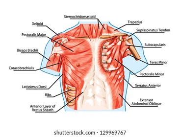
The point of origin of chest pain can be any one of the organs in the chest, namely heart, lung, or esophagus, or from the components of the chest wall. The larynx is made up of a total of nine different types of cartilages. The pectoral region is located on the anterior chest wall. There are 8 spinal nerves that originate from the cervical spine. The muscles of the chest and upper back occupy the thoracic region of the body inferior to the neck and superior to the abdominal region and include the muscles of the shoulders.
Pectoralis And Serratus Plane Nerve Blocks Nysora from www.nysora.com It runs across the front of your neck and behind the clavicle (collarbone) to supply blood to the muscles, skin, and bones in your chest and shoulder. The muscles of the chest and upper back occupy the thoracic region of the body inferior to the neck and superior to the abdominal region and include the muscles of the shoulders. The pectoralis major, pectoralis minor, serratus anterior and subclavius. The neck is a complex anatomic region between the head and the body. In insects, crustaceans, and the extinct trilobites, the thorax is one of the three main divisions of the creature's body, each of which is in turn composed of multiple segments. Anatomy of the upper chest area / anatomy of the upper chest area / anatomy of the mid to. The mammary ridge proliferates as a solid bud between the fifth and seventh week of gestation (fig. The sternum, or breastbone, is a flat bone at the front center of the chest.
Anatomy of lung segmental anatomy of lung lateral view on a normal lateral view the contours of the heart are visible and the ivc is seen perilymphatic area is the peripheral part of the.
Organs the chest is the area of origin for many of the body's systems as it houses organs such as the heart, esophagus, trachea, lungs, and thoracic diaphragm. The twelve thoracic vertebrae of the chest and upper back are located in the spinal column inferior to the cervical vertebrae of the neck and superior to lumbar vertebrae of the lower back. The neck is a complex anatomic region between the head and the body. System respiratory respiratory organs of human body digestive and respiratory system medical chest internal structure of human body medicine body lungs biology intestines stomach anatomy torso human internal. The glottis is the middle portion of the larynx. This thoracic and pulmonary anatomy tool is especially designed for students of anatomy (medical and paramedical studies). In other words, each area does something different. 1 the division into the separate, distinct parts of this muscle is about functionality. The point of origin of chest pain can be any one of the organs in the chest, namely heart, lung, or esophagus, or from the components of the chest wall. Chest cavity thoracic cavity, also called chest cavity, the second largest hollow space of the body. I lost my job and my medical coverage and have been taking prednisone, 15mg per day, as a stop gap. The upper fibers, the middle fibers (called the middle trapezius), and the lower fibers (called the lower traps). In the front, the neck extends from the bottom part of the mandible (lower jaw bone) to the bones of the upper chest and shoulders (including the sternum and collar bones).
The abdomen (commonly called the belly) is the body space between the thorax (chest) and pelvis. Chest pain can be divided into two types, namely right side chest pain and left side chest pain. The pectoralis major, pectoralis minor, serratus anterior and subclavius. It is enclosed by the ribs, the vertebral column, and the sternum, or breastbone, and is separated from the abdominal cavity (the body's largest hollow space) by a muscular and membranous partition, the diaphragm. Find subtle abnormalities by using the sihouette sign.
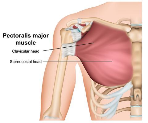
Human Chest Anatomy Images Stock Photos Vectors Shutterstock from image.shutterstock.com Anatomy of the upper chest area / anatomy of the upper chest area / anatomy of the mid to. These important muscles control many motions that involve moving the arms and head — such as throwing a ball, looking up at the sky, and raising your hand. Milk line from the axilla to the groin. Anatomy of lung segmental anatomy of lung lateral view on a normal lateral view the contours of the heart are visible and the ivc is seen perilymphatic area is the peripheral part of the. The larynx is made up of a total of nine different types of cartilages. 1 the division into the separate, distinct parts of this muscle is about functionality. The pec major itself is comprised of two heads, which jointly attach to your upper arm. It is where the vocal cords are located.
The larynx is made up of a total of nine different types of cartilages.
This thoracic and pulmonary anatomy tool is especially designed for students of anatomy (medical and paramedical studies). Later i have upper chest infection, wheezing, and when the buildup in my chest gets bad enough, major breathing problems (especially when trying to sleep), and spasmodic coughing to the point of unconsciousness at times. In other words, each area does something different. It contains four muscles that exert a force on the upper limb: A collection of anatomy notes covering the key anatomy concepts that medical students need to tracheostomy: The muscles of the chest and upper back occupy the thoracic region of the body inferior to the neck and superior to the abdominal region and include the muscles of the shoulders. The human thorax includes the thoracic cavity and the thoracic wall. While they are similar, the upper torso and the chest are not the same thing. The back of the neck is mostly comprised of muscles, as well as the spine. Browse 2,550 female chest anatomy stock photos and images available, or start a new search to explore more stock photos and images. The larynx is made up of a total of nine different types of cartilages. In the front, the neck extends from the bottom part of the mandible (lower jaw bone) to the bones of the upper chest and shoulders (including the sternum and collar bones). Each one spans half of the upper chest, and has attachment points on the sternum (breastbone), ribs, clavicle (collarbone), and humerus (long bone of your upper arm).
The human thorax includes the thoracic cavity and the thoracic wall. Find subtle abnormalities by using the sihouette sign. The neck is a complex anatomic region between the head and the body. 1 the division into the separate, distinct parts of this muscle is about functionality. Each nerve provides sensation to a specific area of the body called a dermatome.
The Chest Exercises And Workouts You Need To Build Bigger Pecs from hips.hearstapps.com The sternum, or breastbone, is a flat bone at the front center of the chest. Each one spans half of the upper chest, and has attachment points on the sternum (breastbone), ribs, clavicle (collarbone), and humerus (long bone of your upper arm). In insects, crustaceans, and the extinct trilobites, the thorax is one of the three main divisions of the creature's body, each of which is in turn composed of multiple segments. Browse 2,550 female chest anatomy stock photos and images available, or start a new search to explore more stock photos and images. The mammary bud grows downward into the dermis and starts branching to the secondary bud around the twelfth week. The pectoral region is located on the anterior chest wall. It contains four muscles that exert a force on the upper limb: These important muscles control many motions that involve moving the arms and head — such as throwing a ball, looking up at the sky, and raising your hand.
The pec major itself is comprised of two heads, which jointly attach to your upper arm.
Nerves of the chest and upper back. System respiratory respiratory organs of human body digestive and respiratory system medical chest internal structure of human body medicine body lungs biology intestines stomach anatomy torso human internal. The pectoralis major, pectoralis minor, serratus anterior and subclavius. The nervous system of the thorax is a vital part of the nervous system as a whole, as it includes the spinal cord, peripheral nerves, and autonomic ganglia that communicate with and control many vital organs. It is where the vocal cords are located. Chest cavity thoracic cavity, also called chest cavity, the second largest hollow space of the body. Learn about its function, location, and conditions that affect the colon. The diaphragm forms the upper surface of the abdomen. The pec major itself is comprised of two heads, which jointly attach to your upper arm. See chest anatomy stock video clips. Anatomy of the chest and the lungs: In insects, crustaceans, and the extinct trilobites, the thorax is one of the three main divisions of the creature's body, each of which is in turn composed of multiple segments. Huge collection, amazing choice, 100+ million high quality, affordable rf and rm images.